A Unified Longitudinal Shape Analysis Framework for Computational Anatomy
By Nikhil Singh, Jacob Hinkle, Sarang Joshi and P. Thomas Fletcher
A key factor in treating diseases can be identification of biomarkers which provide objective measures of disease progress. This article, based on an ISBI 2013 Best Student Paper award, reports on progress in identifying an imaged biomarker for Alzheimer’s disease.
It is well known that the human brain undergoes continuous and gradual structural changes in anatomy with aging. In cognitive disorders, such as the Alzheimer’s disease, the brain attenuation typically occurs at a much accelerated rate as compared to that in normal aging. Alzheimer’s disease is a senile dementia characterized by severe behavioural, cognitive and functional impairment accompanied by distinctive neuroanatomical shape changes. A major obstacle in conducting clinical trials for treatments of Alzheimer’s disease is the enormous cost. This is compounded by the fact that no standardized quantitative imaging-based biomarkers yet exist. Imaging-based biomarkers hold the potential to reduce the costs of clinical trials by the quantitative monitoring of disease progression and treatment effects. Longitudinal quantitative imaging biomarkers hold the promise of identifying prodromal disease, enabling the trials to more efficiently recruit patients most likely to be impacted by treatment and providing earlier detection of outcomes.
The primary goal of our research is to build longitudinal models on a population of multiple individuals repeatedly scanned in time. The aim of our longitudinal models is to quantify the biomarker trajectories and their statistical relationship to cognitive decline. We have developed manifold-based longitudinal statistical models that will inherently accommodate the nonlinearity of anatomical changes associated with the progression of the disease.
The special nature of longitudinal or repeated, time-series data of individual subjects requires the development of novel approaches for image analysis in order to tackle the challenging issues of multiple image registration, segmentation, detection, etc., in the presence of dynamic geometry and contrast. Our research provides methodologies and tools to overcome limitations of the current practice of independent analyses of repeated data, in order to enable researchers to extract and model dynamic properties which otherwise would not become accessible. An important component of this research is to extend commonly known longitudinal analysis concepts for scalar data to statistical analysis of high-dimensional 4D image data, shape and appearance changes related to functional properties.
Characterizing individual neurodegeneration: In our research, the first question we address is the modeling of individual continuous trajectory of atrophy of whole brain in 3D along time [2].

Figure 1: Six years predicted future brain atrophy for an individual with Alzheimer’s disease. Geodesic regression estimates smooth continuous trend of whole brain anatomy changes for an individual. This subject had 4 MRI scans within a period of two years (70-72 years). The last two columns show the predicted brain generated for ages 74 and 76.
Figure 1 shows this trajectory for an individual diagnosed with Alzheimer’s disease at baseline. This individual had MRI scans taken for two years starting at baseline age of 70. Our geodesic regression algorithm captures the estimates of the smooth trend of atrophy. The extrapolation along this trajectory of atrophy can predict the future trend of atrophy for this Alzheimer’s subject. The mathematical formulation of vector momenta based diffeomorphic evolution of 3D shape enables us to evolve the brain to 6 years in the future. The resulting generated brains exhibit a clear trend in shrinking hippocampus, and expanding ventricles along with cerebro spinal fluid across the whole brain with time. These patterns of atrophy are well known to characterize the disease progression in AD.
Modeling population 4D evolution: The second goal of our research is to build a full longitudinal model on a population of multiple individuals repeatedly scanned in time. There are several technological challenges to such analyses. First, while univariate or low-dimensional mixed-effects models are well-developed in statistics literature, little work exists for analyzing high-dimensional longitudinal data such as that derived from medical images. Analysis of high-dimensional data is not as simple as applying univariate longitudinal models to each variable separately. Such a point-by-point analysis fails to take the covariance of the different anatomical brain regions into account, which results in poor estimates of the group trend. In this research we have developed methods for modeling the inherent nonlinearity of anatomical shape changes using differential geometric manifold based methods.
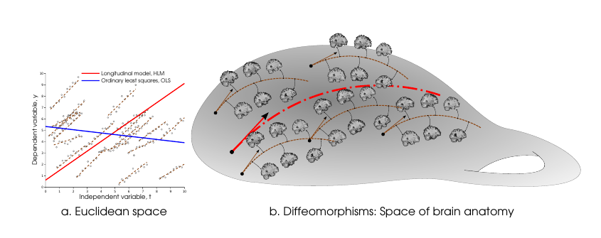
The most common approach is to apply mixed-effects models, pioneered by [6]. These models are hierarchical and also called hierarchical linear models (HLMs), characterizing each individual trend as a linear model built upon a linear model of the overall population trend. The population trends are modeled as “fixed effects”, i.e., unobserved parameters, while the individual trends are modeled as “random effects”, which themselves are random variables with certain distributional parameters. Figure 2 (a) shows a comparison of a regression model (that ignores the fact that the data is longitudinal) compared with a longitudinal model. It can be seen that the longitudinal hierarchical model correctly takes into account the grouping of data into individuals with repeated measurements. This results in the group trend (fixed effects) better reflecting how individuals progress on average. The shape of the regression curve does not match individual trajectories.

Figure 2: Longitudinal modeling via Hierarchical Geodesic Model (HGM).
We have extended this idea to develop hierarchical models for analyzing lon- gitudinal data on infinite-dimensional shape spaces of diffeomorphisms [1]. In this research, we have proposed a novel hierarchical geodesic model (HGM), which generalizes HLMs to the manifold setting (Figure 2 b). Our proposed model explains the longitudinal trends in shapes represented as elements of the group of diffeomorphisms. The individual level geodesics represent the trajectory of shape changes within individuals. The group level geodesic represents the average trajectory of shape changes for the population. The methodological tools developed as a part of this work enable researchers to extract and model dynamic properties from repeated medical image scans. We expect these models will change the outlook of the medical imaging research community towards the longitudinal analysis of high-dimensional information available from medical images.
For Further Reading
1. N. Singh, J. Hinkle, S. Joshi, P. T. Fletcher, “A Hierarchical Geodesic Model for Diffeomorphic Longitudinal Shape Analysis”, Information Processing in Medical Imaging (IPMI) (June 2013)
2. N. Singh, J. Hinkle, S. Joshi, P. T. Fletcher, “A vector momenta formulation of diffeomorphisms for improved geodesic regression and atlas construction”, International Symposium on Biomedial Imaging (ISBI) (April 2013)
3. N. Singh, P. T. Fletcher, J. S. Preston, R. D. King, J. S. Marron, M. W. Weiner, and S. Joshi, “Quantifying anatomical shape variations in neurological disorders,” Medical Image Analysis. Submitted.
4. N. Singh, A. Wang, P. Sankaranarayanan, P. Fletcher, S. Joshi, 2012. “Genetic, structural and functional imaging biomarkers for early detection of conversion from mci to ad.”, Ayache, N., Delingette, H., Golland, P., Mori, K. (Eds.), Medical Image Computing and Computer-Assisted Intervention MICCAI 2012. Vol. 7510 of Lecture Notes in Computer Science. Springer Berlin Heidelberg, pp. 132-140.
5. N. Singh, P. T. Fletcher, J. S. Preston, L. Ha, R. King, J. S. Marron, M. Wiener, S. Joshi, 2010. “Multivariate statistical analysis of deformation momenta relating anatomical shape to neuropsychological measures. “, Proceedings of the 13th international conference on Medical image computing and computer-assisted intervention: Part III. MICCAI’10. Springer-Verlag, Berlin, Heidelberg, pp. 529-537.
6. N. M. Laird, J. H. Ware. “Random-effects models for longitudinal data.”, Biometrics 38(4), pp. 963-974 (1982)
Contributor
 Nikhil Singh is pursuing his PhD in computer science at the University of Utah. He works at the Scientific Computing and Imaging Institute (SCI) with Dr. Tom Fletcher and Dr. Sarang Joshi as a research assistant. The focus of his research is on statistical shape analysis. He develops geometric models that represent human brain anatomy and its variability to model neurodegeneration associated to aging and cognitive disease progression. Read more


 Nitish V. Thakor is is a Professor of Biomedical Engineering at Johns Hopkins University, Baltimore, USA, as well as the Director of the newly formed institute for neurotechnology, SiNAPSE, at the National University of Singapore.
  Max Viergever is Professor and Head of the Department of Medical Imaging at Utrecht University. He also holds appointments as Professor in Physics and Computer Science at Utrecht University.
  Michael Liebling is an assistant professor in the Department of Electrical and Computer Engineering at the University of California, Santa Barbara. He received his PhD from the Ecole Polytechnique Federale de Lausanne (EPFL), Switzerland.
  Lawrence Staib received his Ph.D. degree in Engineering and Applied Science from Yale University, New Haven, CT. He is now Associate Professor of Biomedical Engineering, Diagnostic Radiology and Electrical Engineering at Yale University.
  Stephen R. Aylward is the founder of and Senior Director of Operations at Kitware's North Carolina office, and an adjunct professor in computer science at UNC. Stephen received a Ph.D. in medical image analysis from UNC.
  Bram van Ginneken is Professor of Functional Image Analysis at Radboud University Nijmegen Medical Centre. He is co-chair of the Diagnostic Image Analysis Group within the Department of Radiology, together with Nico Karssemeijer.
  Emily L. Dennis has a B.A. degree in Psychology from Whitman College ('08) and is currently pursuing her Ph.D. in Neuroscience at UCLA with Dr. Paul Thompson. There she researches brain structural and functional connectivity - specifically, how they change over development, are influenced by genetics, and how they are related to cognitive function.
  Nikhil Singh is pursuing his PhD in computer science at the University of Utah. He works at the Scientific Computing and Imaging Institute (SCI) with Dr. Tom Fletcher and Dr. Sarang Joshi as a research assistant.