Systematic Design of a Cross-Polarized Dermoscope for Visual Inspection and Digital Imaging
By Hening Wang, Xin Xu, Xiaoqin Li, Peng Xi, and Qiushi Ren
NOTE: This is an overview of the article, which appeared in the December 2011 issue of the IEEE Instrumentation & Measurement Magazine.
Click here to read the article.
A dermoscope is a diagnostic device that can image the skin in situ and is used for early diagnosis of melanoma and pigmented skin lesions. In this paper, we describe the design and construction of a cross-polarized dermoscope including the illumination evaluation, imaging design, and the mechanical setup. By using the cross-polarization dermoscope, specular reflection from the superficial layer of the skin is largely eliminated. Therefore, deeper layers of the skin, such as the inner pigments and the capillary blood vessels, can be visualized.
Introduction
Skin, as the largest organ of our body, plays an important role in protecting deeper tissues from damage by the external environment. The incidence of skin cancer is increasing each year; therefore, tools to examine skin lesions are of great importance. Early detection of skin cancers leads to effective treatment and thus a high cure rate. However, common clinical diagnostic methods, such as biopsy-pathological analyses, are restricted in the case of dermal examinations due to their invasive nature.
The authors explain that while it is difficult to see deeper layers of tissue because of the shielding effect of the upper layers, polarized light has been used to help assess boundaries of skin cancers. Because of the light scattering nature of biological tissues, the polarization angle is altered for scattering caused by the deeper layers, while the superficial back scattered light maintains its polarization status.
This paper describes the cross-polarization dermoscope, an instrument that can be used to obtain dermal images from the deep layers of the skin. The aim of the cross-polarization dermoscope is to aid the physician to diagnose conditions such as melanoma with a deeper penetration depth imaging. When used to evaluate melanomas and other pigmented skin lesions, the dermoscope magnifies a pigmented lesion and allows the dermatologist to see through the stratum corneum, thereby permitting a detailed view of the deep structural information of the skin.

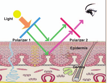
Fig.1. Schematic diagram of the principles of a
cross-polarization dermoscope
System Design
The principle of a cross-polarization dermoscope is shown in Fig. 1. Cross-polarization detection is realized through the application of a pair of polarizers placed perpendicularly between the light source and the observer. The incident light passes through Polarizer 1 and illuminates the skin. Because the specular reflection from the surface maintains the same polarization as the incident light, it will be blocked by Polarizer 2 whose polarization direction is perpendicular to the incident light; however, the scattering of the light from the deep tissue structures alters its polarization, so that it can partially pass through the second polarizer for imaging.
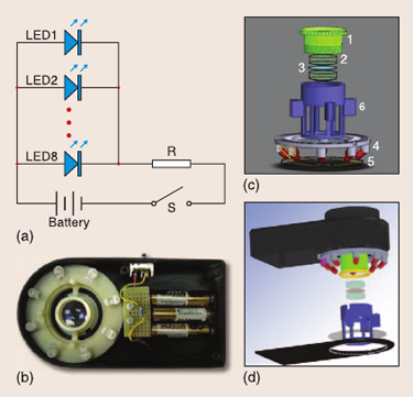
The paper describes simulations that aided the design of the three main parts of the dermoscope – the illumination array, the imaging lenses, and the mechanical assembly (Fig. 2). Refer to the paper for descriptions of the simulations, including a link for details of the mechanical and optical simulations.

Fig. 2. Circuit and mechanical design of the dermoscope: (a) circuit design, (b) the dermoscope system prototype, (c) an illustration of the mechanical design and main components of the optical system, and (d) the CAD assembly of the dermoscope.
Results
The authors used the finished dermoscope to observe human pigmentation lesions on the skin surface and captured the images using a digital camera. They demonstrate how the polarization scheme allows the improved visualization of the edges of a mole on the skin, and the identification of capillaries, which aid in the medical diagnosis. They also state that the diagnostic capabilities of the device could be improved by the use of spectral illumination and detection, in place of the white light illumination they employed.
ABOUT THE AUTHORS
Hening Wang received her B.S. degree in Biomedical Engineering from Shanghai Jiao Tong University in 2010. She is currently a Ph.D. candidate in Dr. Peng Xi’s research group at Peking University, where her research is on the application of nanomaterials in optical microscopy.
Xin Xu received her B.S. degree in Biomedical Engineering from Shanghai Jiao Tong University in 2009. She is currently a Ph.D. candidate in the School of Biomedical Engineering, Science and Health Systems at Drexel University, where her research is on the use of piezoelectric cantilevers for cancer detection.
Xiaoqin Li received his B.Sc. degree in Biomedical Engineering from Shanghai Jiao Tong University in 2011. He is currently a Ph.D. candidate in the biomedical engineering system research group at the University of North Carolina at Charlotte, where his research is on 3-D Human motion analysis and biomechanics.
Peng Xi (xipeng@coe.pku.edu.cn) received a Ph.D. degree in Optical Engineering from Shanghai Institute of Optics and Fine Mechanics, Chinese Academy of Sciences in 2003. He is currently an Associate Professor at Peking University. His research interests include biomedical optics, nonlinear optical microscopy, and optical nanoscopy.
Qiushi Ren received his Ph.D. Degree in 1990 at Ohio State University. Dr. Qiushi Ren became a faculty member of the Department of Biomedical Engineering with an adjunct appointment in the Department of Ophthalmology and Bascom Palmer Eye Institute of University Miami before joining the University of California, Irvine. He then returned to China and the Department of Biomedical Engineering at Shanghai Jiao Tong University and is currently the Head of the Department of Biomedical Engineering, College of Engineering, at Peking University.


