Feeling the Pressure
By Jeffrey Chun-Hui Lin, Yu Zhao, Po-Jui Chen, Mark Humayun, and Yu-Chong Tai
NOTE: This is an overview of the entire article, which appeared in the September 2012 issue of the IEEE Nanotechnology Magazine.
Click here to read the entire article.
Accurate measurement of Intraocular Pressure, or IOP (the fluid pressure inside the eye) is important for the diagnosis and treatment of human eye diseases, such as glaucoma.
Currently, IOP is clinically determined with an external tonometer which measures the deflection of the cornea in response to either direct physical contact or a puff of air. Obviously these methods do not lend themselves to continual monitoring of IOP. However, it is desirable to perform continual monitoring on patients with elevated pressure, as the IOP can change significantly over time due to daily activities.
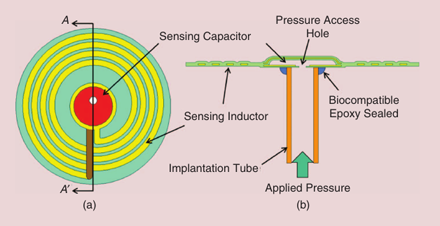
The authors describe a number of active and passive implantable pressure sensors that have been reported in the literature. Their lab has developed a passive implantable sensor, consisting of an integrated series (variable) capacitor, an inductor and a resistor. The resonant frquency of the circuit, when implanted, varies with the intraocular pressure. An external oscillator circuit (perhaps mounted on an eyeglass frame) detects the resonant frequency.

The new IOP sensor design: (a) top view of the sensing part and
(b) AA0 cross-section view of the IOP sensor.
The paper describes the theory, design, and testing of this device. They determined that, with a properly designed high-resolution external coil reader, the sensor can resolve pressure differences of <1mm Hg. By way of comparison, the IOP of a normal human eye is about 10 mmHg, and the maority of glaucoma patients exhibit an IOP of over 20 mmHg.

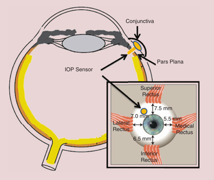
The newly designed IOP sensor is implanted at the pars plana with the implantation tube going through the choroid while the sensing part still remains outside the choroid but under the conjunctiva of the eye.
The authors plan a surgical placement of the device as shown above. The resulting sensor can allow continuous and wireless IOP monitoring.
ABOUT THE AUTHORS
Note: More information on the authors is given in the complete article.
Jeffrey Chun-Hui Lin (linch@mems.caltech.edu) received his B.S. degree in mechanical engineering from National Taiwan University, Taipei, in 1999, and his M.S. and Ph.D. degrees in electrical engineering from the California Institute of Technology (Caltech), Pasadena, in 2007 and 2012, respectively. He is currently working at MiniPumps, LLC in Pasadena, California, as a microdevice and quality control engineer.
Yu Zhao (yzhao@mems.caltech.edu) received her B.S. (Hons) degree in electrical engineering from Peking University in 2008 and her M.S. degree from the California Institute of Technology in 2009. She is currently a Ph.D. candidate in Prof. Y.C. Tai’s group, focusing on developing wireless power transmission solutions for retinal and spinal cord prosthesis devices.
Po-Jui Chen (pojui.chen@gmail.com) received his B.S. degree in mechanical engineering from National Taiwan University, Taipei, in 2002, and his M.S. and Ph.D. degrees in electrical engineering from the California Institute of Technology (Caltech), Pasadena, in 2004 and 2008, respectively. He has been active in technology research and development, mainly in the area of MEMS.
Mark Humayun (MHumayun@doheny.org) received his B.S. degree from Georgetown University, Washington, District of Columbia, in 1984, his M.D. degree from Duke University, Durham, North Carolina, in 1989, and his Ph.D. degree from the University of North Carolina, Chapel Hill, in 1994. He finished his training by completing an ophthalmology residency at Duke University and a fellowship in vitreoretinal diseases at Johns Hopkins Hospital. He is currently a professor of ophthalmology, biomedical engineering, and cell and neurobiology at the University of Southern California, Los Angeles, where he is the director of the National Science Foundation Biomimetic MicroElectronic Systems Engineering Research Center. He is also the director of the Department of Energy Artificial Retina Project that is a consortium of five Department of Energy laboratories and four universities, as well as industry.
Yu-Chong Tai (yctai@its.caltech.edu) received his B.S. degree from National Taiwan University and his M.S. and Ph.D. degrees in electrical engineering from the University of California, Berkeley, in 1986 and 1989, respectively. He is currently a professor of electrical engineering, mechanical engineering, and bioengineering at the California Institute of Technology (Caltech), Pasadena. His main research interest at Caltech is MEMS for biomedical applications, including lab-on-a-chip and neural implants.


