
Standardization of Three Dimensional (3D) and Four Dimensional (4D) Based Medical Application
By Young Lae Moon, Dae Ok Kim, Wonbong Lim
Standardization of medical 3D and 4D Application has not been pioneered.
Additive manufacturing, otherwise known as medical 3D, is driving major innovations in many areas, such as manufacturing, engineering, art, education and medicine. Especially, the medical field is greatly becoming interested in this technology with the ability to create solutions specifically tailored towards the patient. From the creation of 3D models that help surgeons plan operations, to the fabrication of patient-specific titanium implants, 3D printing is already changing the traditional medical industry. In our Working group, sponsored by IEEE Engineering in Medicine and Biology Society (EMBS) as a primary sponsor for 3D Based Medical Application Working Group (EMB/Stds Com/3333.2) with the Computer Society as joint sponsor, practical applications of medical 3D has been suggested the demand for technical standards for clinical educational utilities.
Medical imaging and modeling procedures for solid organ 3D printing.
Medical images from hospitals consist of a two-dimensional (2D) dataset and provide human body information as a slice, however the human body has a three-dimensional (3D) morphology. If we reproduce a 3D morphology via simulations, we might be able to obtain more information about the body as well as contribute in the clinical environment to both better treatment and surgical outcomes. The objective for solid organ 3D printing is to generate 3D medical data from 2D images. Although doctors spend a great deal of time and effort in this process, the resultant 3D data are usually different in each institute. A standardized procedure provides standard, simple and accurate 3D data for solid 3D printing.
The procedure for hard and soft tissue 3D printing
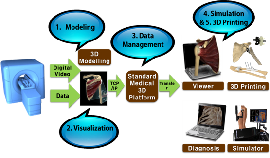
Figure 1. The Procedure for hard and soft tissue 3D printing
Standardization in hard and soft tissue printing involves the use of medical scanning devices to acquire physical data models with density and size characteristics which are necessary to develop comparative analysis data. In order to achieve an accurate segmentation, it is necessary to apply certain segmentation algorithms, including processing step, such as extracting bone features with image enhancement and density selection. The standard for hard and soft tissue 3D printing defines a procedure that increases the precision of 3D printing model output of hard or soft tissues in medical images. In addition, medical imaging and modeling procedures for hard and soft tissue 3D printing will include the following features: 1) Modeling for image enhancement, 2) Visualization in medical image, 3) Data management, 4) Simulation and 5) 3D printing (Fig.1).
Standardization of personalized artificial joint implant 3D model design
The goal of medical 3D printing in the orthopedic field is to reproduce the normal biomedical functions of missing bones. It is necessary to put and apply the artificial joint replacement as the presently feasible intermediate step. This standard is to apply the output to the operation by individually optimizing the shape of the implants of the lost joint based on the rotation data of the positional rotation of the mirrored motion in the normal joint. The use of CAD based on medical image is essential, and a designing technique that minimizes the modeling error is needed. Therefore, definition of optimal design elements for medical 3D printing and development of technical standards based on the analysis of medical elements of artificial joint output are required for analysis of patient’s three-dimensional model data, artificial joint template and other technical factors. In order to maximize the patients and physician’s satisfaction with implant surgery, the accuracy of artificial prosthesis placement is important and surgical guide model design techniques are required to minimize errors.
Standard for in vivo evaluation of three-dimensional printed polymeric scaffolds in bone defects
The standard specifies the in vivo experimentation required for the biological assessment of three-dimensional (3D0 bioprinted polymeric scaffolds intended for the use in bone regeneration. 2D bioprinted scaffolds are gaining increasing attention, and animal experiments are fundamental in assessing their performance prior to potential clinical use. This international standard can be applied to the preclinical assessment such as animal experiments to evaluate the in vivo performance of 3D bioprinted porous polymeric scaffolds.
More recently, the 3D medical applications working group (P3333.2 WG) added 5 project authorization requests (PARs). The approved PARs are: “Standard for Soft Tissue Modeling for Medical 3D Printing,” “Standard for Hard Tissue Modeling for 3D Printing,” “Standard for Surgical Guide Design Modeling for Medical 3D Printing,” “Standard for Artificial Joint Implant Design Modeling for Medical 3D Printing,” and “Standard for In Vivo Evaluation of 3D Printed Polymeric scaffolds in bone defects.” The resulting family of standards under IEEE P3333.2.5 (Standard for Bio-CAD file Format for Medical Three-Dimensional (3D) Printing) will create a substantial basis for improved medical diagnoses, surgical simulations, implant design, tissue engineering and virtual endoscopy, and personalized medical services.
 Young Lae Moon
Chairman & Professor
Orthopaedic Department
Chosun University Hospital
ylm2103@gmail.com