Development of the “rich club” in brain connectivity networks from 438 adolescents & adults aged 12 to 30
By Emily L. Dennis, Neda Jahanshad, Arthur W. Toga, Katie L. McMahon, Greig I. de Zubicaray, Ian B. Hickie, Margaret J. Wright, Paul M. Thompson
Rapid advances are being made in studies of the brain’s connections, which use neuroimaging techniques and mathematical algorithms to analyze anatomical and functional networks. The ‘rich club’ is a central core of highly interconnected nodes, that are critical for network communication. The rich club has been studied as a property of brain networks, but no one has yet examined how it matures in the developing brain. Here we examine the developmental trajectory of rich club organization in anatomical brain networks between ages 12 and 30. These concepts may be advantageous for studying brain maturation and abnormal brain development.
The ‘rich club’ is the central core of a network, made up of high degree nodes that are highly interconnected – more so than would be expected simply given their high degree. The rich club nodes and edges have a large impact on the network’s efficiency, as they participate in a disproportionately high number of the shortest paths. This is a recent concept in brain connectomics, and no one had yet researched the developmental trajectory of the rich club organization, as the brain matures. Given this, and its central importance to network communication, we chose to examine how the rich club coefficient – and which nodes were included in the rich club – changed across adolescence and early adulthood.
The rich club coefficient describes the density of connections between rich club nodes, simply calculated by examining the connections that exist compared to the number of connections possible between rich club nodes. This coefficient is typically normalized based on the properties of a set of simulated random networks, a step that is necessary to determine whether or not rich club organization exists. We simulated 50 random networks, of the same size and degree distribution, but with edges distributed randomly. The rich club coefficient is then normalized relative to its mean value in these networks. A normalized rich club coefficient greater than 1 indicates rich club organization. We ran our statistics, however, on the unnormalized rich club coefficient, as it reflects the true density of connections between these nodes.
Our study was conducted in 438 subjects between the ages of 12 and 30 who are part of a 5-year project examining healthy twins in Australia. Subjects were scanned with HARDI (high angular resolution diffusion imaging) and T1-weighted anatomical scans. We ran tractography on the HARDI data to generate 68×68 matrices reflecting the fiber density between 34 cortical labels per hemisphere. Age effects on the rich club coefficient were estimated using the general linear mixed effects model, including random effects to account for kinship in our sample, and including age, sex, and intracranial volume as covariates.
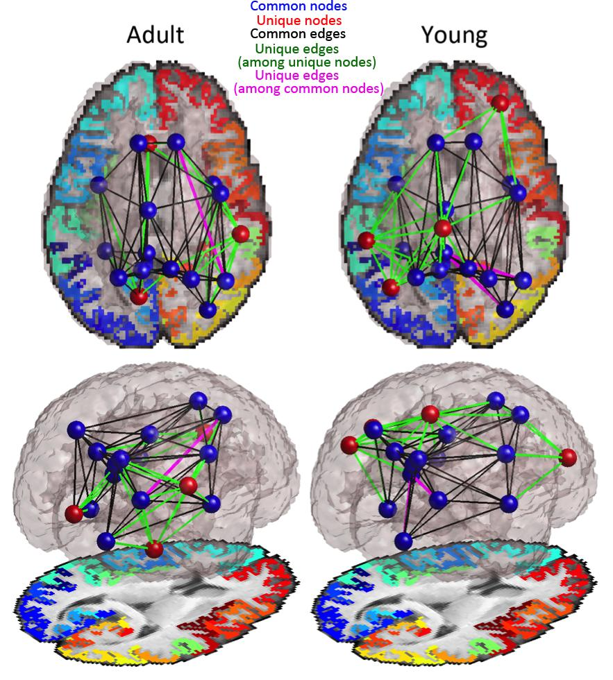
We found a significant age effect on the rich club coefficient, indicating that the density of connections between rich club nodes increases with age. We also looked at which nodes were included in the rich club. Examining statistically determined high degree nodes (degree more than 1 standard deviation above mean), we found 11 nodes in the rich clubs of our ‘adult’ and ‘young’ groups, 2 of which were unique to either group (Figure 1). For this analysis, the adults and the young groups had slightly different degree cut-offs for rich club membership, due to differences in average degree between groups. These cut-offs were statistically determined, but we were curious how the groups would compare at the same degree threshold. Another analysis at a slightly lower degree threshold yielded 18 nodes in rich club, 4 of which were unique to either group (Figure 2). To determine whether these differences could be due to sampling, we split the adults into 2 random groups and examined differences in rich club membership between the groups, across 20 trials. Using the statistically determined criteria for rich club membership, the average difference in rich club nodes across all 20 trials between groups was 0 nodes. This tells us that the core is remarkably stable, and that our results are in fact true age effects.

Figure 1: Rich club networks in young (12 and 16 year old) and adult (20-30 year old) cohorts at statistically determined cut-offs for rich club membership (young=19, adult=20). Edges are thresholded to only show those present in >75% of subjects in either group.

Figure 2: Rich club networks in young (12 and 16 year old) and adult (20-30 year old) cohorts at a degree threshold of 17, both groups still had the same size rich club. Edges are thresholded to only show those present in >75% of subjects in either group.
The areas that were included in the rich club were largely frontal and parietal areas, with a few others, similar to the original paper on this topic. These regions are important for a very broad range of functions such as executive control, sensory processing, and emotion, to name just a few. One existing hypothesis is that the rich club serves to connect areas of various intrinsic connectivity networks to facilitate communication and switching between them. An intrinsic connectivity network (ICN) is made up of regions of the brain whose activity is temporally synchronous. A number of ICNs have been reliably detected across subjects. The rich club includes regions from each of the major ICNs, so this is a possibility. The nodes that were unique to each group were too few to draw conclusions from, but the fact that they are different is interesting. The rich club is a stable core of the network. Yet even at this later stage of brain maturation, we see significant restructuring of the core. Establishing the developmental trajectory of these brain connectivity metrics in healthy individuals is a first step towards determining how and when children with neurodevelopmental disorders may deviate from this trajectory, and which interventions might help them return to the normal trajectory.
Contributor
 Emily L. Dennis has a B.A. degree in Psychology from Whitman College (’08) and is currently pursuing her Ph.D. in Neuroscience at UCLA with Dr. Paul Thompson. There she researches brain structural and functional connectivity – specifically, how they change over development, are influenced by genetics, and how they are related to cognitive function. She worked previously with Dr. Fumiko Hoeft, in the Center for Interdisciplinary Brain Sciences Research at Stanford University, and with Dr. Ian Gotlib, in the Mood and Anxiety Disorders Laboratory, also at Stanford University. Read more


 Nitish V. Thakor is is a Professor of Biomedical Engineering at Johns Hopkins University, Baltimore, USA, as well as the Director of the newly formed institute for neurotechnology, SiNAPSE, at the National University of Singapore.
  Max Viergever is Professor and Head of the Department of Medical Imaging at Utrecht University. He also holds appointments as Professor in Physics and Computer Science at Utrecht University.
  Michael Liebling is an assistant professor in the Department of Electrical and Computer Engineering at the University of California, Santa Barbara. He received his PhD from the Ecole Polytechnique Federale de Lausanne (EPFL), Switzerland.
  Lawrence Staib received his Ph.D. degree in Engineering and Applied Science from Yale University, New Haven, CT. He is now Associate Professor of Biomedical Engineering, Diagnostic Radiology and Electrical Engineering at Yale University.
  Stephen R. Aylward is the founder of and Senior Director of Operations at Kitware's North Carolina office, and an adjunct professor in computer science at UNC. Stephen received a Ph.D. in medical image analysis from UNC.
  Bram van Ginneken is Professor of Functional Image Analysis at Radboud University Nijmegen Medical Centre. He is co-chair of the Diagnostic Image Analysis Group within the Department of Radiology, together with Nico Karssemeijer.
  Emily L. Dennis has a B.A. degree in Psychology from Whitman College ('08) and is currently pursuing her Ph.D. in Neuroscience at UCLA with Dr. Paul Thompson. There she researches brain structural and functional connectivity - specifically, how they change over development, are influenced by genetics, and how they are related to cognitive function.
  Nikhil Singh is pursuing his PhD in computer science at the University of Utah. He works at the Scientific Computing and Imaging Institute (SCI) with Dr. Tom Fletcher and Dr. Sarang Joshi as a research assistant.