Photonics for Life
By Rinaldo Cubeddu, Andrea Bassi, Daniela Comelli, Sergio Cova, Andrea Farina, Massimo Ghioni, Ivan Rech, Antonio Pifferi, Lorenzo Spinelli, Paola Taroni, Alessandro Torricelli, Alberto Tosi, Gianluca Valentini, and Franco Zappa
Many biological mechanisms are related to light interaction or can be evaluated through processes involving energy exchange with photons. Optics has always been a precious tool to evaluate molecular and cellular mechanisms, but the discovery of lasers opened new pathways of interactions of light with biological matter, pushing an impressive development for both therapeutic and diagnostic applications in biomedicine. The recent use of photons in the field of telecommunications has pushed the technology toward low-cost, compact, and efficient devices, making them available for many other applications, including those related to biology and medicine where these requirements are of particular relevance. Moreover, basic sciences such as physics, chemistry, mathematics, and electronics have recognized the interdisciplinary need of biomedical science and are translating the most advanced researches into these fields. The Politecnico school has pioneered many of them, and this article reviews the state of the art of biomedical research at the Politecnico in the field internationally known as biophotonics.
Skin Tumor Detection by Fluorescence Lifetime Imaging Basic Principles and Protocol
Fluorescence techniques for tumor detection can take advantage of endogenous fluorophores, widely present in biological tissues, or can rely on specific exogenous markers with suitable fluorescent properties. Sensitizers used for the photodynamic therapy of tumors (PDT) combine tumor-localizing properties and strong fluorescence. For these reasons, besides being used for therapy, they offer valuable opportunities for tumor detection by fluorescence imaging.
Fluorescence lifetime imaging (FLIM) relies on a series of time-gated images taken a few nanoseconds after the short laser pulses are used to cause the fluorescence. A linear regression allows one to calculate the spatial map of the average decay time as a function of position. Along this line, the authors are performing a clinical study on the detection of skin lesions by means of lifetime imaging of ALA-induced PpIX fluorescence. A challenging goal of this investigation is the progressive reduction in ALA dose, starting from the one used for therapy (20%). This is in agreement with the minimization of undesired skin photosensitization lasting after the diagnostic session [1]. A considerable engineering effort has been spent to assemble a portable instrument for FLIM, starting from a laboratory prototype.
The basic principles of lifetime imaging and the technical details of the instrument are described, together with its preliminary clinical application to the localization of skin lesions. For this study, 16 dermatological lesions in 12 patients have been considered; five basal cell carcinomas (BCCs), two squamous cell carcinomas (SCCs) and nine benign lesions.
The classification of the lesions was performed on the basis of the fluorescence lifetime that resulted from the accumulation of PpIX and the contemporaneous reduction of the endogenous fluorescence in malignant lesions [2]. Both these effects lead to a markedly long-living fluorescence in tumors. The diagnostic assessment of the lesions was performed with histopathologic analysis. The result was a correct classification in five of seven malignancies (with two false negatives) and nine of nine benigns. The present study demonstrates the effectiveness of FLIM, in combination with ALA-induced fluorescence, to detect nonmelanotic skin tumors even at very low dose of the marker [2].
Diffuse Optical Spectroscopy
Basic Principles and Instrumentation
The diffusive nature of most biological tissues has long hindered any extensive measurement of their optical properties, despite the increasing demand for in vivo tissue optical characterization, both for diagnostic and therapeutic purposes. In the wavelength range extending from 600 to 1,100 nm, the reduced scattering coefficient (m’s) is predominant over the absorption coefficient (ma) and cannot be neglected. The study of photon propagation through a diffusive medium in the time domain allows us to disentangle absorption from scattering contributions and to recover information from a few centimeter depth beneath the surface or through 5-6 cm of thickness. More than 20 years of research on this topic has been pioneered at the Department of Physics of Politecnico di Milano, with the development and continuous upgrade of a unique workstation for diffuse optical spectroscopy operated in the time domain [3].
Application Examples
The system was used to explore new potential applications in view of a possible clinical finalization. We performed the first in vivo measurements of the absorption spectrum of photosensitizer drugs used in PDT, demonstrating spectral changes due to the environment [4], and a possible improvement of the irradiating procedure.
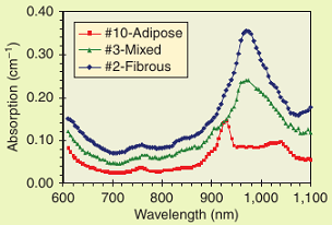
An emerging field was the study of the optical properties of the female breast, with the hope of developing an optical technique for the early diagnosis of breast cancer, as discussed in the following section. Also, the first optical characterization of collagen powder in this spectral range has shown the possibility to devise an optical tool for the assessment of breast cancer risk [5]. Quite recently, increased collagen content was found to be related to a higher risk, and its noninvasive detection could permit earlier diagnosis and more effective prevention [6]. Figure 1 shows an example of in vivo absorption measurements on healthy volunteers with three different breast types. The fibrous breast (associated with a higher mammographic density and thus a higher risk) has a clearly distinguishable spectral pattern. The system was also used to characterize different tissues and components for a better understanding of light propagation into biological media.
Figure 1. Absorption spectra measured in vivo from the breast of three healthy volunteers with different breast types. The fibrous pattern (associated with a highter risk factor) can be easily identified by the larger water absorption peak at 970 nm (blue line).
Basic Principles and Instrumentation
In recent years, both in the United States and Europe, optical mammography has been investigated as a possible means for the diagnosis of breast cancer. Despite the lower spatial resolution as compared to X-rays, optical mammography performed at several wavelengths can couple with a single noninvasive diagnostic tool, which has the ability for lesion localization and a high potential for lesion identification and, more generally, tissue characterization. To take advantage of the potential differences in blood parameters as well as water, lipid, and collagen content between healthy and diseased tissue, we developed a time-resolved optical mammograph designed to collect projection images in compressed breast geometry. In the last version of the instrument, time-resolved transmittance measurements are performed at seven wavelengths using picosecond pulsed diode lasers and two personal computer (PC) boards for time-correlated single photon counting.
As for the lesion detection and characterization, a retrospective study was performed to define the optical appearance of different breast lesions and structures, with the final aim of determining effective diagnostic criteria [7].
Photon-Counting SPAD Array Detector Systems
In recent years, there has been a growing interest in monolithic arrays of SPADs for parallel detection of faint and/or ultrafast optical signals. Parallel detection can lead to a significant reduction of the acquisition time in high-sensitivity measurements. Furthermore, the availability of compact monolithic array detectors can open the way to a further miniaturization of array-based analytical systems.
A System for Fast and Sensitive Analysis of Protein Microarrays
At the Dipartimento di Elettronica e Infomazione, Politecnico di Milano, by using the dedicated SPAD technology, we fabricated several SPAD arrays with large area elements. As an example, a 6×8 SPAD array was developed for chemiluminescent array detection and parallel fluorescence correlation spectroscopy (FCS) [24].
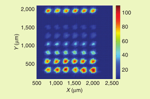
Each pixel of the SPAD array is hybrid connected to an integrated active-quenching circuit (i-AQC) that provides active-quench and active-reset pulses. Starting from this detector, we developed a novel compact instrument capable of ultrasensitive analysis of luminescence originating from multiple proteins in a customized protein microarray. The optical system creates an image of the microarray on a focal plane, where the six by eight SPAD array is located. A digitally controlled motion unit performs the alignment between the protein microarray and the SPAD array. A compact and low-cost field programmable gate array (FPGA) was employed for implementing the counting section. Finally, a communication block provides a simple universal serial bus (USB) interface with the PC for an easy control of the system. The final apparatus is very compact, having a size of 25 x 15 x 15 cm3. Figure 2 shows the image of an array of rabbit IgG detected by incubation with an anti-rabbit IgG labeled with HRP as obtained by the scanning procedure outlined above. The concentration of the immobilized protein ranges from 0.3 to 0.01 mg/mL, and the image was obtained in a total time of only 40 s, i.e., with a 0.2 s\integration time for each pixel of the image. These results advancements are demonstrated not only in production of optoelectronic system for protein analysis but also with the development of the surface chemistry involved in protein immobilization.
Figure 2. Analysis in scanning mode of an array of rabbit IgG detected by incubation with an anti-Rabbit IgG labeled with hepatoma-derived growth factor (HDGF)-related proteins (HRPs). Different concentrations of protein were spotted according to the following scheme: 0.3 mg/mL, negative control (0 mg/mL), 0.01, 0.015, 0.03, 0.05, 0.1, and 0.3 mg/mL.
High-Throughput Fluorescence Correlation Spectroscopy System
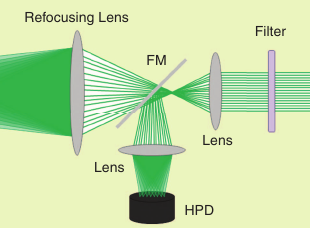
We developed a single-chip array of smart pixels, laid out in 32 rows by 32 columns made in a standard high-voltage 0.35-mm CMOS technology. Every pixel comprises a 20-mm diameter SPAD, a front-end electronics and a processing circuitry for counting photons. Such an array makes it possible to observe binding interactions or the diffusion of fluorescent particles simultaneously in different locations and to observe fast-evolving dynamic systems. We exploit a liquid crystal on silicon (LCOS) to generate a pattern of excitation spots (whose spot number, size and distance can be easily adjusted) and the 32×32 SPAD array to detect the fluorescence light of the emission path (see Figure 3).
Figure 3. Optical setup: LCOS, microscope, and SPAD array.
Conclusions
The short descriptions of the research activities in biophotonics currently active at the Politecnico of Milan make evident that this field has acquired a wide range of applications. Being a technical university, we can benefit from the presence of multidisciplinary competences that are required to promote the research in different application areas. Many of these have already been investigated, but new interesting applications are coming out and will be exploited in the near future.
Acknowledgments
The authors acknowledge the essential contribution of the clinical partners to both the development and evaluation of the clinical applications: Gianfranco Canti (Department of Pharmacology, University of Milan), Gian Maria Danesini (Casa di Cura San Pio X, Milano), Francesca Abbate, Anna Villa, and Enrico Cassano, (Istituto Europeo di Oncologia, Milano).
Rinaldo Cubeddu ( ),
Andrea Bassi (andrea.bassi@fisi.polimi.it),
Daniela Comelli (daniela.comelli@polimi.it),
Andrea Farina (andrea.farina@polimi.it),
Antonio Pifferi (antonio.pifferi@polimi.it),
Paola Taroni (paola.taroni@polimi.it),
Alessandro Torricelli (alessandro.torricelli@polimi.it), and
Gianluca Valentini (gianluca.valentini@polimi.it)
are with the Dipartimento di Fisica, Politecnico di Milano, Milano.
Sergio Cova (sergio.cova@polimi.it),
Massimo Ghioni (massimo.ghioni@polimi.it),
Ivan Rech (ivan.rech@polimi.it),
Alberto Tosi (alberto.tosi@polimi.it),
and Franco Zappa (franco.zappa@polimi.it),
are with the Dipartimento di Elettronica e Informazione, Politecnico di Milano.
Lorenzo Spinelli (lorenzo.spinelli@polimi.it)
is with the Istituto di Fotonica e Nanotecnologie (IFN) – CNR, Milano.
Click here to read the full paper, which appeared in the May/June 2011 issue of the IEEE Pulse magazine.
- R. Cubeddu, A. Pifferi, P. Taroni, P. Taroni, A. Torricelli, F. Rinaldi, and E. Sorbellini, “Fluorescence lifetime imaging: An application to the detection of skin tumors,” IEEE J. Select. Topics Quantum Electron., vol. 5, no. 6, pp. 23-29, 1999.
- R. Cubeddu, D. Comelli, C. D’Andrea, P. Taroni, and G. Valentini, “Time-resolved fluorescence imaging in biology and medicine,” J. Phys. D: Appl. Phys., vol. 35, no. 1, pp. 1-16, 2002.
- A. Pifferi, A. Torricelli, P. Taroni, D. Comelli, A. Bassi, and R. Cubeddu, “Fully automated time domain spectrometer for the absorption and scattering characterization of diffusive media,” Rev. Sci. Instrum., vol. 78, no. 5, pp. 053103-1 – 053103-10, 2007.
- R. Cubeddu, G. Canti, M. Musolino, A. Pifferi, P. Taroni, and G. Valentini, “In vivo absorption spectrum of disulphonated aluminum phthalocyanine in a murine tumor model,” J. Photochem. Photobiol. B, vol. 34, no. 2-3, pp. 229-235, 1996.
- P. Taroni, A. Bassi, D. Comelli, A. Farina, R. Cubeddu, and A. Pifferi, “Diffuse optical spectroscopy of breast extended to 1100 nm,” J. Biomed. Opt., vol. 14, no. 5, pp. 054030-1 – 054030-7, 2009.
- J. Couzin, “Dissecting a hidden breast cancer risk,” Science, vol. 309, no. 5741, pp. 1664-1666, 2005.
- L. Spinelli, A. Torricelli, A. Pifferi, P. Taroni, G. Danesini, and R. Cubeddu, “Characterisation of female breast lesions from multiwavelength time-resolved optical mammography,” Phys. Med. Biol., vol. 50, no. 11, pp. 2489-2502, 2005.


