Electromagnetic Tomography for Medical and Industrial Applications: Challenges and Opportunities
By H.-Y. Wei and M. Soleimani
NOTE: This is an overview of the entire article, which appeared in the March 2013 issue of the Proceedings of the IEEE.
Click here to read the entire article.
This Point Of View article reports on research underway to make “Magnetic Induction Tomography (MIT) a tool for use in medical and industrial applications. As opposed to the familiar “CT Scan”, which uses x-rays to produce images of the body, MIT employs magnetic fields. MIT is a technique that uses electromagnetic fields to examine the passive electromagnetic property (PEP) variation of a material, which can potentially be used for many biomedical applications, such as brain diagnostics. This article reviews the latest progress of MIT, and its way forward into future.
Two of the most widely used tomography techniques that currently exist in the field are X-ray and γ-ray tomography, the so-called “hard-field” tomography. The costs related to hard-field tomography are generally very high, and these systems contain radioactive sources which may cause side effects in patients under long-term exposure.
On the other hand, “soft-field tomography” techniques are also available for use in medical and industrial fields. The difference between the two techniques is that in hard-field tomography the transmitting signal follows a straight line pattern. In soft-field tomography, the transmitting field does not follow the straight line pattern, and the signal distribution depends on the type of the excitation source. A major challenge in the development of MIT is in the dispersed nature of the electromagnetic field (no longer follows the straight line pattern) because the change in conductivity cannot be “localized” and can cause signal, perturbation on any measurement set.

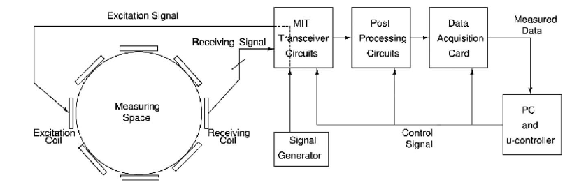
Fig. 1. A complete MIT system block diagram.
Fig. 1 illustrates a typical MIT system, which generally consists of the following:
- an array of evenly spaced coils that are arranged around the measuring object;
- the front-end electronics, for example, receiver buffers, driving amplifiers, and channel multiplexing switches;
- a data acquisition unit;
- a computer and the algorithm which can transfer the signals into MIT images.
The article describes the complexity of the calculations required to produce an MIT image. The authors have constructed several such systems and have tested them in both medical and industrial applications.
The authors believe that a portable, low-cost battery operate MIT system could be developed, and could facilitate medical diagnosis in rural areas. They envision early-stage diagnosis of physiological brain disorders using this tool. They point out the need for further interdisciplinary research.
ABOUT THE AUTHORS
H.-Y. Wei and M. Soleimani are with the Engineering Tomography Laboratory, Department of Electronic and Electrical Engineering, University of Bath, Bath, U.K.
