
IEEE Life Sciences: Spotlight on Research in Biological Imaging
By Cynthia Weber
IEEE Life Sciences spoke with Michael Liebling from the Idiap Research Institute in Switzerland and the University of California Santa Barbara about current research and developments in biological imaging and analysis.
IEEE Life Sciences: What developments in the field of biological image acquisition and analysis are most exciting?
Michael Liebling: There have been several recent breakthroughs in optical microscopy. Some of these have been recognized through the 2014 Nobel Prize in Chemistry, awarded to Betzig, Hell, and Moerner. To me, an especially important aspect was that for at least one of these award-winning techniques, the computational aspect is central: the image is produced via computation. More generally, images are increasingly used to measure and quantify biological processes, not just to qualitatively observe them. Image processing is therefore gaining a more central role in experiment planning in biology and is no longer just used as a last resort tool to salvage poor images.
IEEE Life Sciences: Tell us about your research.
ML: My group develops computational imaging techniques to study biological systems. Concretely, we devise new microscopy protocols and hardware devices, which, when combined with the image processing algorithms we develop, produce images that could not be obtained directly with off-the-shelf microscopes. One example is imaging the beating heart inside developing animal embryos: no commercial microscope can currently deliver 3D volumes at sufficient frame-rate (100–1000 volumes per second). To solve this problem, we have developed methods that combine movies of 2D sections taken at multiple depths and directions to reconstruct a full view of the beating heart. Several collaborating groups now use our tools. Other topics include methods to achieve temporal super-resolution (increase the frame-rate of movies) or deconvolution of blurred microscopy images.
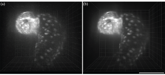
Live 2.5 dpf Tg(cmlc2:eGFP) zebrafish embryo imaged in fluorescence at 60 frames per second. (a) 125 z-slices are synchronized to reconstruct a 3D volume. Due to the low illumination intensity and the short integration time used during acquisition, the resulting image is severely corrupted by Poisson-type noise. (b) Our temporal superresolution reconstruction is able to simultaneously temporally superresolve the image sequence and remove much of the noise. We used nine cardiac cycles with a to reconstruct a single denoised heart beat with an effective sampling rate of 120 frames per second. Scale bar is 100 µm. (Figure adapted from: K. G. Chan, S. J. Streichan, L. A. Trinh and M. Liebling, “Simultaneous Temporal Superresolution and Denoising for Cardiac Fluorescence Microscopy,” in IEEE Transactions on Computational Imaging, vol. 2, no. 3, pp. 348-358, Sept. 2016. doi: 10.1109/TCI.2016.2579606)
IEEE Life Sciences: What is most challenging about your research?
ML Going from an idea that looks promising on paper or even in simulations on synthetic data to implementing a full system that actually performs in practice and can be used by our collaborators is probably the biggest challenge. At the same time, being able to control the entire imaging pipeline offers many opportunities. Sometimes, going back and adjusting the imaging protocol can greatly simplify the subsequent image processing. In my lab we have the possibility to do end-to-end experiments and students will often prepare the biological sample, carry out the imaging under the microscope, then apply the image processing tools they have implemented. While this often requires learning skills in orthogonal topics (biology, optics, signal processing, hardware tinkering) it ends up paying off. Again, image processing is no longer just an “after-thought” but is truly becoming an integral part in the experiment planning stage.
IEEE Life Sciences: What draws you to this type of research?
ML: I like the idea that engineers can contribute to advancing scientific discovery. Interacting with biologists is particularly rewarding; their research is fast-paced and many of them are open to trying out new technology. Their studies also increasingly require quantitative characterization of images. And of course, nothing beats seeing a nice image (or better, a nice movie) of a biological process that hadn’t been observed before.
IEEE Life Sciences: Where do you see the field moving in the next five years?
ML: I think that computational imaging methods will keep gaining importance and acceptance among both industry (for example, by default, many commercial microscopes now serve images only after the raw measurements have undergone processing) and scientists, as the precise modeling of the imaging process, inherent to most computational imaging methods, is also essential for quantitative imaging. We can expect to observe structures and processes at increasingly fine space- and time- scales, buried deep inside organisms.
 Michael Liebling received a M.S. in physics in 2000 and Ph.D. in 2004 both from Ecole Polytechnique Fédérale de Lausanne, Switzerland. From 2004 to 2007, he was a postdoctoral scholar at the California Institute of Technology, Pasadena. In 2007, he joined the faculty of the Department of Electrical and Computer Engineering, the University of California, Santa Barbara, first as an Assistant Professor and, since July 2013, as an Associate Professor. In 2015, he joined the Idiap Research Institute, Martigny, Switzerland, as a Senior Researcher, where he leads the computational bioimaging group. His research interests include biological microscopy and image processing for the study of dynamic biological processes. Dr. Liebling is a Member of the IEEE Signal Processing Society’s Bio-Imaging and Signal Processing Technical Committee, which he chaired in 2014–2015. He serves as an Associate Editor (2015–2016) and Senior Associate Editor for IEEE Signal Processing Letters (2016–2018) and was Technical Program Co-chair of the IEEE International Symposium on Biomedical Imaging in 2011 and 2013. He is the vice-chair of the IEEE Life Sciences Technical Community Steering Committee.