
Medical Imaging in Five Dimensions: Volumetric Color Hearing in Real Time
by Subhamoy Mandal
Multi-Spectral Optoacoustic Tomography (MSOT), a new technique developed based on the principle of multi-wavelength optoacoustics, is capable of high resolution three dimensional (3D) visualizations of molecular probes located deep in scattering living tissues, with resolution and speed representative of ultrasound. This method can simultaneously deliver anatomical, functional, and molecular information with both high resolution and greater penetration depths, and when combined with speed and multi-spectral capabilities, opens the door to imaging in five dimensions.
“It is a very extraordinary sensation to hear a beam of sunlight laugh, cough, and sing, and talk to you with articulate words; but when you understand how these results are accomplished, you see the operation is a simple one,” observes Prof. Alexander Graham Bell during discussion of his famous 1880 Boston lecture, “Upon the production and reproduction of light.”
This discovery of the “photophone” led to the foundation of modern optoacoustic (also called as photoacoustic) imaging, which uses ultrashort laser pulses to generate the signal in tissue chromophores and record it with help of ultrasonic detectors [1]. The journey from the photophone to the first images of animal tissues took nearly a century (see the history of evolution in Figure 1), however, what followed next was a rapid expansion of the field, leading to a new imaging modality offering high-resolution visualization of optical contrast, overcoming the longstanding limitations imposed by light scattering in deep tissues [2].
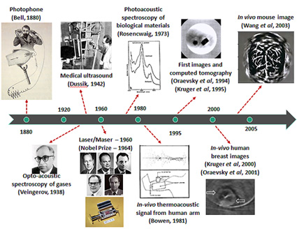
Figure 1. History of development of optoacoustic imaging. Courtesy of Mandal et al., IEEE Pulse (forthcoming, May/June 2015).
Over the last few decades researchers have experimented with different modes and configurations for acquiring better optoacoustic images, succeeding in producing higher resolution images deeper in the tissues [3]. “The true performance of optoacoustic imaging techniques can only be exploited when excitation at multiple wavelengths is used in order to enable highly sensitive spectral differentiation of intrinsic biomarkers and extrinsically administered contrast agents,” Daniel Razansky, Professor of Molecular Imaging at the Technische Universität München, explains. “By detecting tiny sound vibrations, resulting from selective absorption of light at multiple wavelengths, multispectral optoacoustic tomography (MSOT) can now hear color in three dimensions.” The newly developed system can deliver volumetric spectrally enriched (color) images at high spatial resolution and in real time from deep tissues in vivo [4].
The principle of MSOT is based on detection of acoustic signals created through the thermoelastic expansion of tissue under influence of light, representative of ultrasound (lower by a few orders of magnitude). These signals are collected in form of a time-of-flight (ToF) data and then reconstructed using tomographic inversion processes to render 2D/3D images. Though several alternative designs exist, the newly developed portable spherical array probe (by Prof. Razansky’s research group), combined with fast wavelength tuning laser, real-time data acquisition, GPU-based volumetric image rendering, and spectral unmixing, has enabled for the first time volumetric real-time spectrally-enriched (5-dimensional) optoacoustic imaging at centimeter scale depths [5,6].
Time (or speed) is one of the most important dimensions that define a living world, but as we aim for higher resolution we lose speed. MSOT, unlike many other imaging modalities, overcomes this challenge by offering fast imaging capabilities. In fact, the researchers have been able to acquire images in vivo at speeds of tens of frames per second. Imaging in 4D (3D + time) has been made possible not only because of advances in scanning geometries and hardware components, but equally because of enhancement of software components, including inversion and image/signal processing method. Combining the capabilities of fast real time imaging with the multi-wavelength probing has enabled isolation of the distribution of the contrast agent.
The high imaging speed of MSOT has made possible artifact-free handheld imaging, visualization of a beating heart, or real-time imaging of perfusion profiles in tumors and internal organs (kidneys, brain, etc.) of small animals. Multi-spectral (or multicolor) imaging confers molecular specificity and thus provides the capability to quantitatively investigate biological conditions such as hypoxia and nutritional gradients as well as cell viability, proliferation and drug response potentials, which is essential in understanding the dynamics of living tissues and disease prognosis and progression. Thus, 5D imaging of in vivo biological samples has opened up a plethora of new opportunities to understand the dynamics and pharmacokinetics of internal organs and disease progression.
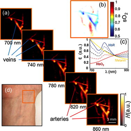
Figure 2. Multispectral tomographic reconstructions of the wrist region. (a) Volumetric (maximum intensity projection along the depth direction) images acquired at 5 different wavelengths in the near-infrared. Two main types of vessels (arteries and veins) can be readily identified by their spectral behavior. (b) Map of blood oxygen saturation, as calculated from images acquired at the different wavelengths. (c) Extinction (absorption) spectra of major tissue chromophores in arbitrary units. The curves for hemoglobin are shown for 100% oxygenation (continuous red line), 75% oxygenation (dashed red line), 50% oxygenation (dashed black line), 25% oxygenation (dashed blue line), and 0% oxygenation (continuous blue line). (d) Color photograph of the imaged region. Image courtesy: Xosé Luís Deán-Ben and Daniel Razansky, Photoacoustics, vol. 1, no. 3, 2014.
MSOT is a technology that is fast maturing and already has taken bold strides towards technology commercialization. As Dr. N. C. Burton, an application specialist with iThera Medical GmbH (Germany), explains, “MSOT has the potential to make a substantial impact on preclinical research as well as on the clinical market. And, finally, though regulatory-approved preclinical devices have already reached the market, medical device permissions in Europe and the United States are still pending. Once approved, multicenter trials in a number of key clinical applications will facilitate the transition of this technology from a highly potent research platform to an accepted medical device.”
Currently, MSOT is being extensively used for small animal imaging trials including in-vivo cell tracking, molecular imaging studies, targeted molecular imaging, as well as functional imaging of the brain/heart, and pharmacokinetic evaluations [7]. Experimental imaging has been initiated for human vasculature imaging (Figure 2), finger imaging for detection of atherosclerosis, ablation studies on cardiac tissues, and breast cancer imaging. However, a long and difficult road exists in front of the research community in optoacoustically visualizing greater depths with enhanced accuracy and contrast. Now with the aid of the newly acquired and powerful capability of extending the paradigm of medical imaging to the fifth dimension, we can hope to reach that destination in the not so distant future.
To learn more about this technology, please watch for an upcoming article in IEEE Pulse (May/June 2015).
References
- D. Razansky, “Multispectral Optoacoustic Tomography—Volumetric Color Hearing in Real Time,” IEEE Journal of Selected Topics in Quantum Electronics, vol. 18, no. 3, pp. 1234-1243, May-June 2012.
- S. Mandal, X. L. Deán-Ben, N. C. Burton, and D. Razansky, “Extending biological imaging to the fifth dimension: Evolution of volumetric small animal multispectral optoacoustic tomography,” IEEE Pulse (in press, 2015).
- V. Ntziachristos, J. Ripoll, L. H. V. Wang, and R. Weissleder, “Looking and listening to light: the evolution of whole-body photonic imaging,” Nat. Biotechnol., vol. 23, pp. 313–320, 2005.
- D. Razansky, M. Distel, C. Vinegoni, R. Ma, N. Perrimon, R. W. Köster, and V. Ntziachristos, “Multispectral opto-acoustic tomography of deep-seated fluorescent proteins in vivo,” Nature Photonics, vol. 3, no. 7, pp. 412-417, 2009.
- X. L. Deán-Ben and D. Razansky, “Functional optoacoustic human angiography with handheld video rate three dimensional scanner,” Photoacoustics, vol. 1, no. 3, pp. 68-73, 2013.
- X. L. Deán-Ben and D. Razansky, “Adding fifth dimension to optoacoustic imaging: Volumetric time-resolved spectrally enriched tomography,” Light: Science & Applications, vol. 3, no. 1, e137, 2014.
- S. Gottschalk, T. F. Fehm, X. L. Deán-Ben, and D. Razansky, “Noninvasive real-time visualization of multiple cerebral hemodynamic parameters in whole mouse brains using five-dimensional optoacoustic tomography,” Journal of Cerebral Blood Flow & Metabolism, 2015.
Contributor
 Subhamoy Mandal is a DAAD Ph.D. scholar with the Institute of Biological and Medical Imaging at the Technische Universität München and Helmholtz Zentrum München. His research focuses on visual quality enhancement in multispectral optoacoustic tomgraphy, and translational molecular imaging applications.
Read more
 Shabana Sayed is a ESHRE certified Senior clinical Embryologist and IVF Laboratory Director at Klinikk Hausken. Her primary areas of expertise are within embryo selection and optimization of laboratory procedures for IVF.
 Subhamoy Mandal is a DAAD Ph.D. scholar with the Institute of Biological and Medical Imaging at the Technische Universität München and Helmholtz Zentrum München. His research focuses on visual quality enhancement in multispectral optoacoustic tomgraphy and translational molecular imaging applications.
 Cristian A. Linte is an Assistant Professor in the Department of Biomedical Engineering and the Chester F. Carlson Center for Imaging Science at Rochester Institute of Technology.
 Ziv Yaniv is a Senior Scientist in the Office of High Performance Computing and Communications at the National Library of Medicine and the National Institutes of Health.