Trends in Bio Imaging and Signal Processing
By Jean-Christophe Olivo-Marin, Laure Blanc-Féraud, Michael Unser, Andrew Laine, and Boudewijn Lelieveldt
The area of bio imaging and signal processing is concerned with the development of dedicated tools for biomedical and bioinformatics applications and with fostering interdisciplinary and integrative approaches of biomedical topics by bridging the biology, medicine and signal processing communities. In-depth understanding of functional and pathological mechanisms in patients or model organisms indeed requires: 1) to visualize and measure in vivo, in a robust and systematic manner their spatiotemporal characteristics and 2) to model the complex signaling networks that regulate the cellular interplay of proteins and gene expression. To that end, increasingly complex imaging devices and image processing protocols are being applied in biological and medical imaging. Similarly, sophisticated bioinformatics methods are used in genomics and proteomics to assemble, decipher and compare data from large-scale sequencing data.
Imaging devices give access to information ranging from the nano to the macro through the micro scale and can visualize, for example, sub-cellular components and processes to investigate cellular dynamics (Fig. 1) or full organs to characterize diseases in animal models (Fig. 2), single patients or patient cohorts. Biomedical scientists are however faced with the problem of visualizing, analyzing and processing the resulting huge volume of data. Image processing scientists have been providing solutions by developing dedicated algorithms: for example in the context of high throughput screening, they have engineered fast, robust algorithms with minimal user interaction that are able to classify, characterize and test the action of thousands of drugs on cells or tissues.
In the area of microscopy, concurrent to major advances in the instrumentation, the capabilities of modern microscopes have been further enhanced with the help of sophisticated signal processing for denoising, deconvolution, or tomographic reconstruction. Several outstanding problems like high levels of noise, photo-bleaching, or the presence of aberrations still remain and are active areas of research.
In medical imaging, the combination of detailed structural imaging modalities such as MR, with molecular imaging can help monitor the biochemical onset of disease and therapy and evaluate structural and functional consequences over time. In longitudinal imaging studies of cohorts, major challenges lie in the sheer data volumes that necessitate full automation, and in feature extraction and data mining of very large image databases. There is also a strong trend towards objective quantitative benchmarking of analysis methods, like in the case of liver, coronary, brain, and carotid segmentation, or image registration.
An important common trend is the design of user-friendly imaging software that can be readily used by practitioners with minimal knowledge of computer science, and can provide a major help in the computer-aided visual interpretation and quantification of highly complex and intertwined data.
Read more on this topic in the article, Trends in Bio Imaging and Signal Processing, which will be published in the November 2011 Issue of IEEE Signal Processing Magazine.
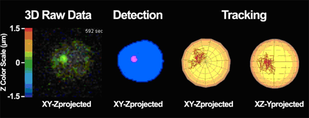
Figure 1: visualization, detection and tracking of a single gene locus in the nucleus of a yeast cell imaged in fluorescence real time confocal microscopy (from Cabal et al., 2006).
![]()
Figure 2: An articulated planar reformatted visualization of a mouse, where the skull has been selected and is shown at three different timepoints in the focus views. The visualization shows a slice of the volume with a corresponding bounding box. Additionally, the global overview shows a semi-transparent surface rendering of the atlas surfaces. From Kok et al, 2010.

![]()
About the authors:
Jean-Christophe Olivo-Marin(jcolivo@pasteur.fr) is a Research Director at Institut Pasteur in Paris
Michael Unser (michael.unser@epfl.ch) is a Professor at Ecole Polytechnique de Lausanne
Laure Blanc-Féraud (Laure.Blanc_Feraud@inria.fr) is a Research Director at CNRS-I3S in Sophia Antipolis
Andrew Laine (laine@columbia.edu) is a Professor at Columbia University in New-York
Boudewijn Lelieveldt (B.P.F.Lelieveldt@lumc.nl) is a Professor at Leiden University in Leiden and at Delft University of Technology in Delft, the Netherlands.
- Cabal, G., Genovesio, A., Rodriguez-Navarro, S., Zimmer, C., Gadal, O., Lesne, A., Buc, H., Feuerbach-Fournier, F., Olivo-Marin, J.-C., Hurt, E.C., and Nehrbass, U. (2006) SAGA interacting factors confine sub-diffusion of transcribed genes to the nuclear envelope, Nature, 441, pp. 770-773
- Kok P, Baiker M, Hendriks EA, Post FH, Dijkstra J, Löwik CW, Lelieveldt BP, Botha CP. Articulated planar reformation for change visualization in small animal imaging. IEEE Transactions on Visualization and Computer Graphics 2010 Nov-Dec;16(6):1396-404.
